Pacemaker Application
Permanent pacemaker
It is used in cases where the event requiring battery implantation is
considered to be permanent. The generator is placed in the chest or abdomen by
creating a pocket under the skin. However, it is usually placed on the left
side of the chest wall. If it is to be placed on the chest wall, patients may
be asked which side they prefer.
How is a pacemaker implanted?
Informed consent is obtained from the patient or their relatives for
pacemaker implantation.
Temporary pacemaker implantation:
Temporary pacemaker implantation is usually performed while patients are
hospitalized for associated heart conditions (e.g. following a heart attack).
The procedure is performed in the patient's room or in the cath lab. After
sedation (if necessary) and local anesthesia, a small sheath is placed in the
neck or groin area. The cable from the generator is passed through this sheath
to the heart. If necessary, x-rays (scopy) are used to ensure that the cable is
placed in the appropriate place in the heart. The pacemaker outside is fixed in
a suitable place. Patients should not touch this unit and should limit their
activities while the temporary pacemaker is in use.
In rare but necessary cases, a temporary pacemaker is connected to the heart
with a needle through the patient's chest wall or, more rarely, through the
esophagus.
Permanent pacemaker implantation:
Permanent pacemaker implantation is a more invasive (bloody) procedure and
is considered minor surgery. Permanent pacemaker implantation can be performed
in a cardiac catheterization laboratory, electrophysiology laboratory, hospital
operating room, or outpatient surgery department.
Pacemaker
In this procedure, the patient's heart rate and blood pressure are
monitored and local anesthetic is given to the patient. The application area is
cleaned and shaved. Depending on where the pacemaker is to be implanted, one of
two methods is used:
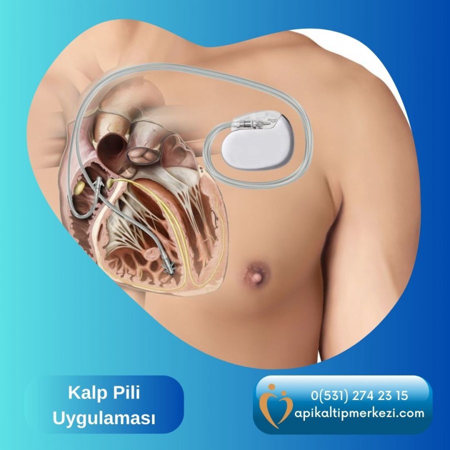
If the pacemaker is to be implanted in the chest wall (endocardial
implantation), a small incision is made just below the collarbone to create a
small surgical pocket. The wires from the generator are passed through a vein
in the upper chest and visually inserted into the right atrium or right
ventricle under X-ray guidance. The end of the cable (electrode) is attached to
the inner surface of the heart with a special screw-shaped tip.
Battery cables (with conductive electrodes at the ends)
If there is more than one cable, the procedure is repeated. The generator
is placed in the pocket opened below the collarbone. After insertion, the skin
is closed with sutures. Thus, nothing of the pacemaker can be seen from the
outside. The whole procedure takes about 1 hour.
Here we see a pacemaker that was previously implanted under the skin under
the collarbone on the right side as a bulge under the skin. In patients with a
high subcutaneous fat layer, this bulge may be absent and the pacemaker may not
be noticed at all.
In a less common procedure known as epicardial implantation (outside of the
heart), the lead is placed on the outer surface of the heart. Using this
method, the surgeon opens the chest wall, the lead is placed on the surface of
the heart and the generator is placed under the skin in the upper abdomen. This
alternative is only used when it is not feasible for the wires to pass through
the veins to reach the inner surface of the heart (e.g. in some congenital
heart diseases or if the patient is a child).
After insertion:
Shortly after implantation, a chest x-ray is taken to check that the device
is properly placed. The pacemaker can be programmed with the programming device
placed on the chest. The patient does not feel anything during this procedure.
Depending on the patient's age and general health,
hospitalization for 1-2 days is recommended after permanent pacemaker
implantation. Recommendations for the appropriate activity level of the patient
in the period immediately after the procedure are explained by the doctor.
Patients should avoid contact sports, heavy lifting, and vigorous movements of
the arm on the side of the pacemaker so that the electrode does not dislodge.
After the suture is closed, there may be some stiffness at the suture site for
a certain period of time. However, the stiffness disappears as the wound heals.
However, any signs of infection (discharge, inflammation of the surgical wound)
should be reported immediately to the doctor who performed the procedure. Until
the suture heals completely (7-10 days), the patient can wash and shower by
paying attention to the suture and drying this area.